2020 Virtual AIChE Annual Meeting
(162ah) Multimodal Probe Development for Specific Mitochondria Imaging with Aggregation-Induced Emission and PET
Authors
Yu, K. - Presenter, Zhejiang University
Xu, Y., Zhejiang University
Zhang, H., The Second Hospital of Zhejiang University
Tian, M., The Second Hospital of Zhejiang University School of Medicine
He, Q., Zhejiang University
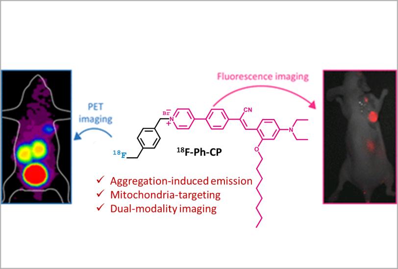
Breast cancer has become the most common malignant tumor in women with rapidly increased incidence rate and younger age of onset. Traditional method for the diagnosis of breast cancer is based on histological analysis which is invasive and inefficient. The emerging molecular imaging techniques provide noninvasive tools for precise and accurate detection of various types of cancer in vivo. However, a lack of imaging probe with high resolution and high specificity has hindered the progress in the early detection of tumor, resulting in poor prognosis after clinical treatments. Mitochondria play critical roles in the development and survival of normal cells and cancer cells. A larger mitochondrial membrane potential in cancer cells make it an important target for imaging probe development. Recently, a number of fluorescent probes with aggregation-induced emission for mitochondria imaging have been reported and exhibited excellent performance in cancer cell imaging. In this work, we have developed a multimodal mitochondria-targeted probe F-Ph-CP with aggregation-induce emission for both fluorescence and PET (positron emission tomography) imaging. F-Ph-CP stains mitochondria of breast cancer cells specifically with high photostability and low cytotoxicity. Then, we could substitute the built-in fluorine atom in F-Ph-CP with radioactive 18F and successfully perform in situ PET imaging for breast cancer, together with fluorescence imaging. The combination of PET and aggregation-induced emission demonstrates the first in vivo visualization of breast cancer by both imaging modality, which shows great potential in preoperative tumor detection and intraoperative optical navigation.


