(23a) Post-Formulation 64-Cu Labeled Nanotracers to Investigate Size and Surface Property Effects on Biodistribution and Tumor Localization in a Mouse Pancreatic Cancer Model
AIChE Annual Meeting
2020
2020 Virtual AIChE Annual Meeting
Particle Technology Forum
Novel Nanoparticles and Nanostructured Materials for Pharmaceuticals and Medical Applications
Monday, November 16, 2020 - 8:00am to 8:15am
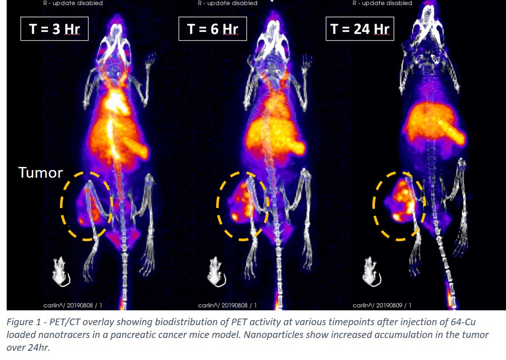
Positron emission tomography (PET) is an extremely sensitive diagnostic tool for use in the early detection of cancers and other diseases. FDG-PET utilizing the PET active glucose analog, 18-fluorodeoxyglucose (18-FDG), has shown promise in clinical cancer imaging but still has several shortcomings. For example, since FDG utilizes glucose metabolism to detect tumors, it is less effective for slow-growing tumors and for differentiating cancers from the surrounding inflammatory tissue. Here we present the formation of nanoparticles using the continuous and scalable flash nanoprecipitation (FNP) process. Phthalocyanine-based dyes are encapsulated inside NPs through a rapid mixing and precipitation process that is stabilized by an amphiphilic block copolymer. After incubation of these NPs with copper ions in solution, the encapsulated dyes exhibit absorbance shifts indicative of the chelation of copper ions into the macrocyclic ring of the phthalocyanine. This absorbance shift was employed to quantify chelation and optimize reaction conditions. Thus, using this same method but instead incubating the NPs with positron-emitting 64-Cu yields PET active nanotracers. Under very mild conditions in a pH 5.5 aqueous buffer, the radiolabeling yield was quantitative after 3hr of incubation at 37C. This technology, when combined with the flexibility of FNP, allows for long timepoint biodistribution tracking of NPs with a variety of different properties such as size and surface charge. In particular, when PS-b-PEG is used as the stabilizing polymer, NPs exhibit long circulation times and enhanced tumor uptake due to the enhanced permeability and retention (EPR) effect. This was demonstrated using our PET nanotracers in a xenograft pancreatic tumor model, which showed high levels of PET activity in circulation at 3hrs and 6hrs post-injection and increasing tumor accumulation over 24hrs (Fig. 1). The development, optimization, and validation of a scalable method to produce nanotracers promises to expand the applications of NP-based PET imaging.