
(572d) Delivery of CRISPR/Cas9 Gene-Editing Systems By Cell-Penetrating Magnetite Vehicles: Synthesis, Characterization and in Vitro Testing
AIChE Annual Meeting
2020
2020 Virtual AIChE Annual Meeting
Nanoscale Science and Engineering Forum
Bionanotechnology for Gene and Drug Delivery I
Wednesday, November 18, 2020 - 8:45am to 9:00am
One of the main challenges in gene therapy is the transport of genetic material into the cells of interest since a number of obstacles need to be overcome including rapid degradation of genetic material by the physiological environment, limited cell uptake and poor endosomal escape. As a result, few alternatives have been implemented to tackle these issues such as the use of viral vectors, and the use of nanostructured materials [5]–[7]. Due to biosafety concerns, using viral vectors has been questioned, while some of the nanostructured materials have shown lower delivery efficiencies [8]–[10].
To improve the efficacy of the delivery nanomaterials capable of responding to magnetic fields are used. This approach is known as magnetofection and relies on using magnetic fields to guide the delivery of the nanovehicles [11]–[14]. Using magnetic nanomaterials has attracted significant attention for applications in biomedicine due to their ease of handling, their ability to transport various types of biomolecules, and the possibility of controlling their fate to specific tissues and organs [15]–[17]. Consequently, the chances for side effects can be significantly reduced [18], [19].
One of the most appealing magnetic nanomaterials is magnetite, which has gained attention over the past decades thanks to its applicability as cell marker, active biologicals separation, drug delivery, and as contrast agent in MRI testing [20]–[23]. This could be attributed to their high biocompatibility and proven possibility of renal excretion as ferritin [21], [24], [25].
To enable the delivery of gene therapies via nanomaterials, the genetic material needs to be covalently conjugated to the surface of the nanomaterials with using highly-reactive functional groups such as thiol, amine, hydroxyl or carboxyl [26]. Additionally, nucleotide cargoes should be able to be released intracellularly and ideally reaching the nucleus. For this purpose, a number of chemistries have been incorporated into the delivery vehicles that respond to environmental changes or stimuli making the system smart [25]. With the emergence of CRISPR/Cas9 systems, more robust and complex interventions can be achieved, and consequently larger nucleotide sequences need to be delivered efficiently, in a cost-effective manner [9], [27].
As abovementioned, the objective of this study is to develop a nanostructured platform for immobilization, and intracellular release of nucleic acids with application in gene therapy. The multifunctional nanostructured carrier (i.e., nanobioconjugate) was based on the surface functionalization of magnetite with an organosilane molecule followed by a Polyether Amine (PEA) or a Polyethylene Glycol (PEG) surface spacer, and finally, a molecule containing a reducible disulfide linker. Simultaneously, a potent translocating, endosome escaping protein was co-immobilized on the surface to increase the efficiency of biologically active genetic material reaching the nuclei. The reducible disulfide linker is formed by direct conjugation of a thiolated 65 bp tag to the free thiol on the surface of the nanobioconjugate, which is complementary to a non-encoding sequence within the delivery vector. Once the nanobioconjugate reaches the intracellular space, the disulfide bond is reduced, and the cargo nucleotide delivered.
Magnetite was synthesized by chemical co-precipitation of the iron chlorides (II) and (III) in a sodium hydroxide 1M solution [28], [29]. Further details of synthesis can be consulted elsewhere [28], [29]. The nanoparticles were subsequently functionalized with the organosilane 3-Amino Propyl Triethoxy-Silane (APTES) to render free amine groups on the surface. The surface spacer PEA (Jeffamine 600) or PEG was conjugated followed by the 3-[(2-aminoethyl)dithio]propionic acid] (AEDP) containing a reducible disulfide bond [16]. The translocating molecule selected for this study was the Outer Membrane Protein A (OmpA) of Escherichia coli due to the recently reported penetrating and endosome escape abilities [28]. To investigate the role of the surface spacer, we prepared three different nanobioconjugates, the first one was produced by directly conjugating the AEDP to the surface of the silanized nanoparticles (OmpA-magnetite-AEDP) (Figure 1a), the second one contained PEG (OmpA-magnetite-PEG-AEDP) (Figure 1b) before AEDP and third one PEA (OmpA-magnetite-PEA-AEDP) (Figure 1c). For amine-amine conjugation, we used glutaraldehyde to form imine bonds, while for amine-carboxyl we used NHS/EDC to form amide bonds. The nanobioconjugates were characterized by FTIR, TGA, TEM, and DLS. The FTIR allowed us to confirm the characteristic bands of the polymer and OmpA. TGA analysis confirmed a conjugation efficiency of PEG of about 1.5% and of PEA of about 5% while for OmpA the efficiency approached 1.5%. TEM allowed us to confirm the typical morphology for the nanoparticles and an average diameter for individual particles of about 15-18 nm. Hemolysis and platelet aggregation exhibited values below 5%.
The delivery of the conjugated material was first tested in vitro with the aid of DTT and GSH as reducing agents. For the experiments, we conjugated BSA and Rhodamine and tracked the time evolution of the delivered molecules via Bradford and spectrofluorimetry, respectively. It appears that the highest release was obtained for the OmpA-magnetite-PEA-AEDP followed by OmpA-magnetite-PEG-AEDP and finally, OmpA-magnetite-AEDP. With these experiments, we decided to deliver to neuroblastome cells to evaluate endosomal escape. This was accomplished by labeling the nanobioconjugates with Rhodamine B (OmpA-magnetite-PEA-AEDP-Rhodamine B). Pearson’s correlation coefficients were calculated to evaluate the colocalization of the nanobioconjugates with the endosomes (labeled with Lysotracker Green). The results suggest an endosomal escape of about 85%. Further evidence of escape and even cytosol distribution was provided by a calculated surface area coverage of about 60%.
Future experiments will be focused on the hybridization of a gene sequence for the expression of the fluorescent protein mCherry. The repair of missing nucleotides will be via homologous recombination using a Gibson Assembly kit. The obtained nanobioconjugate will be delivered to cells to evaluate transfection efficiencies. Detailed cytotoxicity experiments will be also required to assure a complete biocompatibility analysis.
[1] Abarca-Barriga, H. H., et. al. “Risk factors in genetic diseases ,†Acta Med Peru, 2018.
[2] RodrÃguez-Yunta, E. “Gene therapy and ethics principles,†Acta Bioeth., 2003.
[3] GarcÃa, A. H., “Gene Therapy with Non-Viral Vectorsâ€, 2015.
[4] Austin-Ward, E. D., et. al. “Gene Therapy and Its Applications,†Rev. Med. Chil., 1998
[5] Y. Weng et al., “Improved Nucleic Acid Therapy with Advanced Nanoscale Biotechnology,†Molecular Therapy - Nucleic Acids, 2020
[6] Penn, S. G., et. al. “Nanoparticles for bioanalysis,†Curr. Opin. Chem. Biol., 2003
[7] Liu, T., et al., “Calcium phosphate nanoparticles as a novel nonviral vector for efficient transfection of DNA in cancer gene therapy,†Cancer Biother. Radiopharm., 2005
[8] Mohammad, A., et. al. “Nonviral Vectors for Gene Therapy,†ScienceDirect, 2015
[9] Lino, C. A., et. al. “Delivering crispr: A review of the challenges and approaches,†Drug Deliv., 2018
[10] Wang, Y., et. al. “Co-delivery of drugs and DNA from cationic core-shell nanoparticles self-assembled from a biodegradable copolymer,†Nat. Mater., 2006
[11] Plank, C., et. al. “Magnetofection: Enhancing and targeting gene delivery with superparamagnetic nanoparticles and magnetic fields,†in Journal of Liposome Research, 2003
[12] Schillinger, U. et al., “Advances in magnetofection - Magnetically guided nucleic acid delivery,†J. Magn. Magn. Mater., 2005
[13] Krötz, F., et. al. “Magnetofection Potentiates Gene Delivery to Cultured Endothelial Cells,†J. Vasc. Res., 2003
[14] Gersting, S. W. et al. “Gene delivery to respiratory epithelial cells by magnetofection,†J. Gene Med., 2004
[15] Wegscheid, M. L., et. al. “The art of attraction: Applications of multifunctional magnetic nanomaterials for malignant glioma,†Expert Opin. Drug Deliv., 2014
[16] Ramanujan, R. V., “Clinical Applications of Magnetic Nanomaterials,†Sch. Mater. Eng.
[17] Kumar, C. S, et. al. “Magnetic nanomaterials for hyperthermia-based therapy and controlled drug delivery,†Adv. Drug Deliv. Rev., 2011
[18] AsÃn Pardo, L. “BIOMEDICAL APPLICATIONS OF MAGNETIC NANOPARTICLES: MAGNETIC HYPERTHERMIA IN DENDRITIC CELLS AND MAGNETOFECTION IN BRAIN CELLS.â€
[19] TARTAJ, P., et al., Biomedical Applications of Magnetic Nanoparticles, 2007
[20] Kratz, H. et al., “MPI Phantom Study with A High-Performing Multicore Tracer Made by Coprecipitation,†Nanomaterials, 2019
[21] Chanana, M., et. al. “Fabrication of Colloidal Stable, Thermosensitive, and Biocompatible Magnetite Nanoparticles and Study of Their Reversible Agglomeration in Aqueous Milieu,†Chem. Mater., 2009
[22] Pan, B.F., et. al. “Dendrimer modified magnetite nanoparticles for protein immobilization,†J. Colloid Interface Sci., 2005
[23] Estrada de la Vega, A., et. al. “Aglomerados de nanopartÃculas basados en magnetita y CTAB,†2013
[24] N. Habibi, “Preparation of biocompatible magnetite-carboxymethyl cellulose nanocomposite: Characterization of nanocomposite by FTIR, XRD, FESEM and TEM,†Spectrochim. Acta - Part A Mol. Biomol. Spectrosc., 2014
[25] Xu, Z. P., et. al. “Inorganic nanoparticles as carriers for efficient cellular delivery,†Chem. Eng. Sci., 2006
[26] Fraile, M. C., “Study of the interactions between nanoparticles of noble metals and DNA,†2016
[27] Hsu, P. D., et. al. “Development and Applications of CRISPR-Cas9 for Genome Engineering,†Cell, 2014.
[28] Lopez-Barbosa, N., et al., “Magnetite–OmpA Nanobioconjugates as Cell-Penetrating Vehicles with Endosomal Escape Abilities,†ACS Biomater. Sci. Eng., 2019.
[29] Cuellar, M. et al., “Novel BUF2-magnetite nanobioconjugates with cell-penetrating abilities,†Int. J. Nanomedicine, 2018.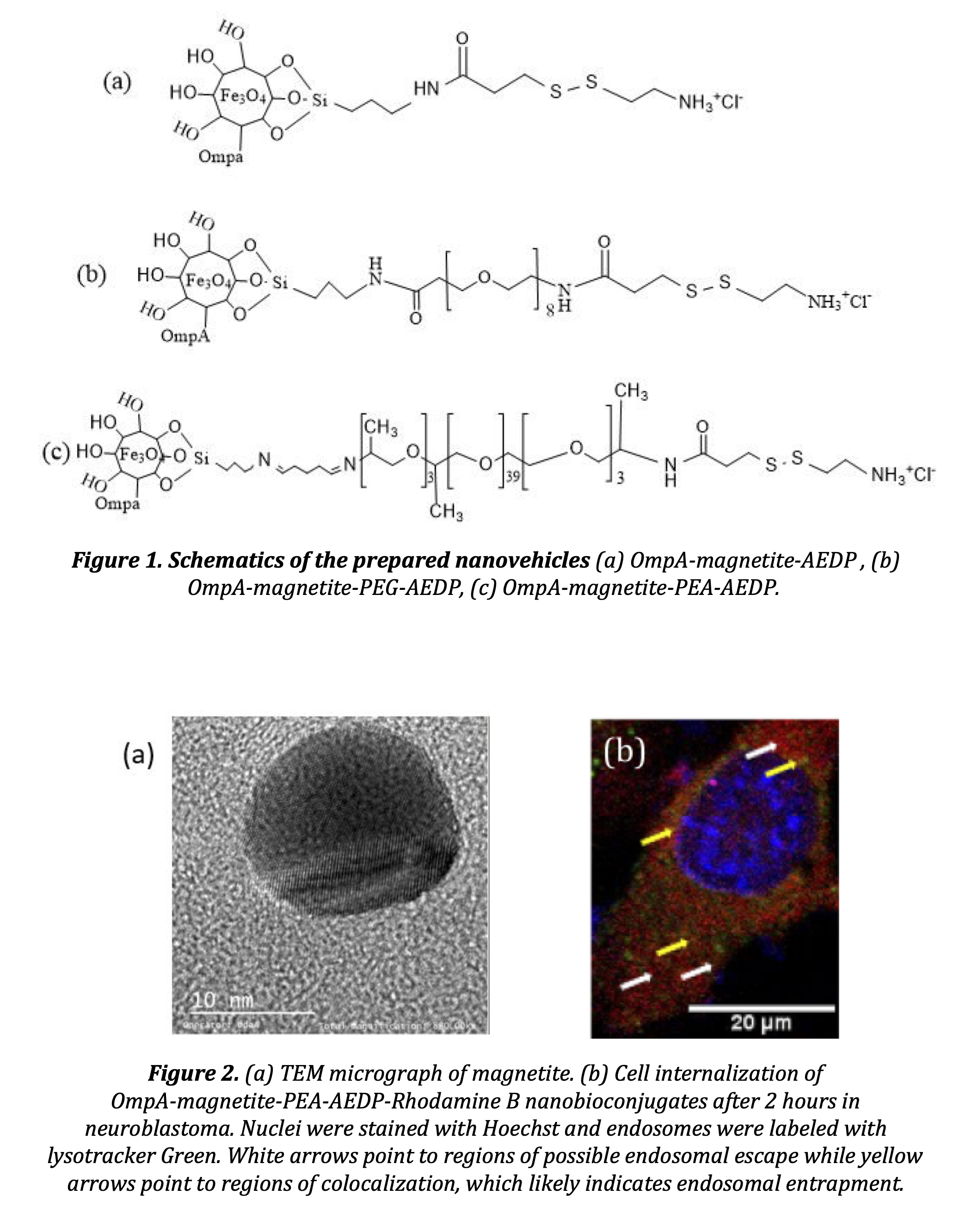
Checkout
This paper has an Extended Abstract file available; you must purchase the conference proceedings to access it.
Do you already own this?
Log In for instructions on accessing this content.
Pricing
Individuals
| AIChE Pro Members | $150.00 |
| AIChE Emeritus Members | $105.00 |
| AIChE Graduate Student Members | Free |
| AIChE Undergraduate Student Members | Free |
| AIChE Explorer Members | $225.00 |
| Non-Members | $225.00 |