(668a) Gelpins: A New Strategy for Discrete Yet Cohesive Tissue Assemblies within ‘Cut & Assemble’ Organ-Chips
AIChE Annual Meeting
2020
2020 Virtual AIChE Annual Meeting
Food, Pharmaceutical & Bioengineering Division
Cells, Organs, and Labs on a Chip: Emerging Applications
Friday, November 20, 2020 - 8:00am to 8:15am
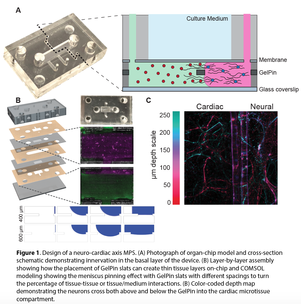
Ensuring contiguous, yet heterogenous structure of multicellular structures is paramount for recapitulating complex microenvironments. For this reason, the innervation of organs where effector neuron bodies are isolated, yet synaptically connected, to the distal organ tissue rarely been demonstrated in vitro. As a proof of principle, GelPins were utilized to establish an MPS of the cardiac sympathetic nervous system incorporating cell laden hydrogels containing both cardiomyocytes (CMs) and adrenergic neurons (Figure 1). Specifically, cardiac cells and neurons were isolated from neonatal rat pups and cultured in situ within a photocrosslinkable gelatin-based hydrogel. Results show neurites extend both above and below the GelPin towards CMs (Figure 1C) after 10 days in culture. Innervation of the cardiac microenvironment was investigated via immunofluorescent imaging at discrete time points throughout the duration of culture and neurite extension quantified using neuron tracing software (Neurolucida®).
The GelPin technology can be additionally leveraged to define boundaries in the z axis by orientating GelPins instead as slats in place of membranes or other means of separating tissue layers (Figure 1B). COMSOL simulations were used a priori confirmed that GelPins with a 1:2 and 1:3 width to spacing ratio (resulting in gaps of 400 and 600 µm, respectively), can function as a pressure barrier in the z-direction. The use of GelPins slats allows tailorable compartment heights down to 10s of microns, optimal for developing culture systems with striated tissue layers like the intestinal lumen, dermis, or retina, while ensuring sufficient diffusion across layers. In summary, this is a promising new approach towards assembling the next generation of MPS for benchtop discovery. Coupled with the ‘cut & assemble’ method, these tools offer robust design flexibility and lower barrier costs to encourage broader use of MPS technology across the scientific community.
1 Hosic, S. et al. Rapid prototyping of a multilayer microphysiological system for primary human intestinal epithelial culture. bioRxiv, 400721 (2018).