(164al) Characterizing a cAMP/Crp Mediated Metabolic Futile Cycle in Bacterial Persisters
AIChE Annual Meeting
2022
2022 Annual Meeting
Food, Pharmaceutical & Bioengineering Division
Poster Session: Bioengineering
Monday, November 14, 2022 - 3:30pm to 5:00pm
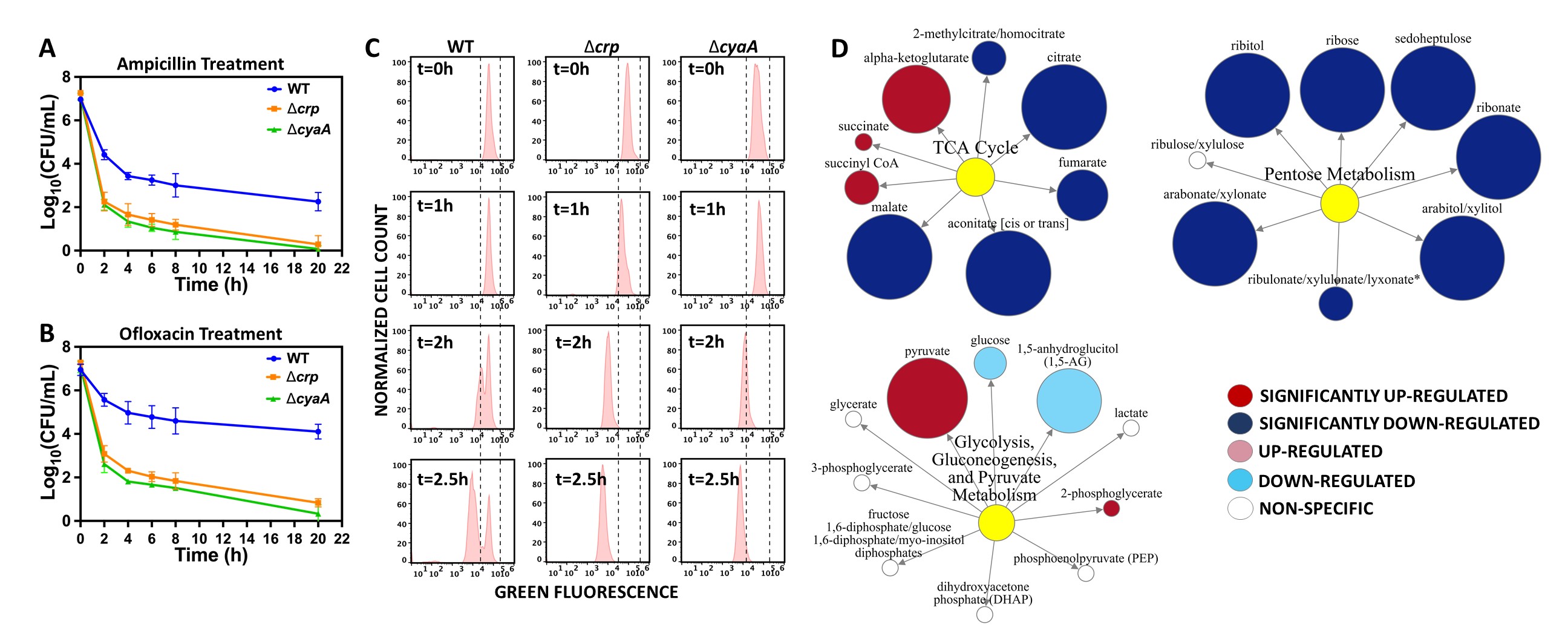
In this study, the deletion of crp (encoding a global transcriptional regulatory protein) and cyaA (encoding adenylate cyclase that synthesizes cAMP) in Escherichia coli reduce type I persister levels significantly compared to wild-type parental strain (Fig. 1A-B). Using an inducible green fluorescent protein (GFP) expression system and protein dilution method, we demonstrated that the crp and cyaA deletions reduce the non-growing cell formation during the stationary phase (Fig. 1C). CRP/cAMP is the DNA-binding transcriptional dual regulator, regulating many genes associated with the central metabolism, so we also performed untargeted mass spectrometry analysis to quantify the metabolites in the wild-type and the mutant crp strain in the stationary phase. The results revealed that metabolites associated with energy metabolism are significantly down regulated in the mutant strain compared to the wild-type (Fig. 1D). When we knocked out some of the genes associated with energy metabolism, we found deletions of these genes also reduce the persistence in E. coli. Overall, with the data presented here, we expect to generate a comprehensive map of the metabolic mechanisms facilitating type I persister cell formation in E. coli.
References
- Balaban, N.Q., Merrin, J., Chait, R., Kowalik, L. and Leibler, S., 2004. Bacterial persistence as a phenotypic switch. Science, 305 (5690), pp.1622-1625.
- Lewis, K. (2007). Persister cells, dormancy and infectious disease. Nature Reviews Microbiology, 5(1), 48-56.
- Lewis, K. (2010). Persister cells. Annual review of microbiology, 64, 357-372.
- Orman, M.A. and Brynildsen, M.P., 2015. Inhibition of stationary phase respiration impairs persister formation in E. coli. Nature communications, 6, p.7983.
Fig. 1: CRP/cAMP-mediated metabolic futile cycle facilitates persister formation in stationary-phase in E. coli (A-B) Persister levels (log-scale) of wild-type (WT) and mutant strains. Late stationary phase cells (i.e., cell populations cultured for 24h) were transferred to fresh LB and treated with ampicillin and ofloxacin to enumerate persisters, as measured by colony-forming units (CFUs). WT persister levels at the late stationary phase were significantly higher than those of mutant strains (N=4). (C) Non-growing cell levels of WT and mutant strains. GFP-positive late stationary-phase cells (harboring an inducible GFP expression system, pQE-80L-gfp) were transferred to fresh media (without inducer) to monitor their proliferation by a fluorescent protein-dilution method. Non-growing cells that did not resuscitate retained their high GFP levels (t=2.5h). A representative biological replicate was shown here. All 3 biological replicates consistently resulted in similar trends. (D) Metabolomics data of the WT and the mutant strain in the late stationary-phase. Red and blue dots represent the metabolites that are significantly upregulated or downregulated in the mutant strain compared to WT, respectively (P-value< 0.05, ANOVA test).