(20c) Directing Multicellular Organization By Varying the Aspect Ratio of Soft Hydrogel Microwells
AIChE Annual Meeting
2022
2022 Annual Meeting
Food, Pharmaceutical & Bioengineering Division
Cell and Tissue Engineering: Mechanical Cues and Cell Behavior
Sunday, November 13, 2022 - 4:06pm to 4:24pm
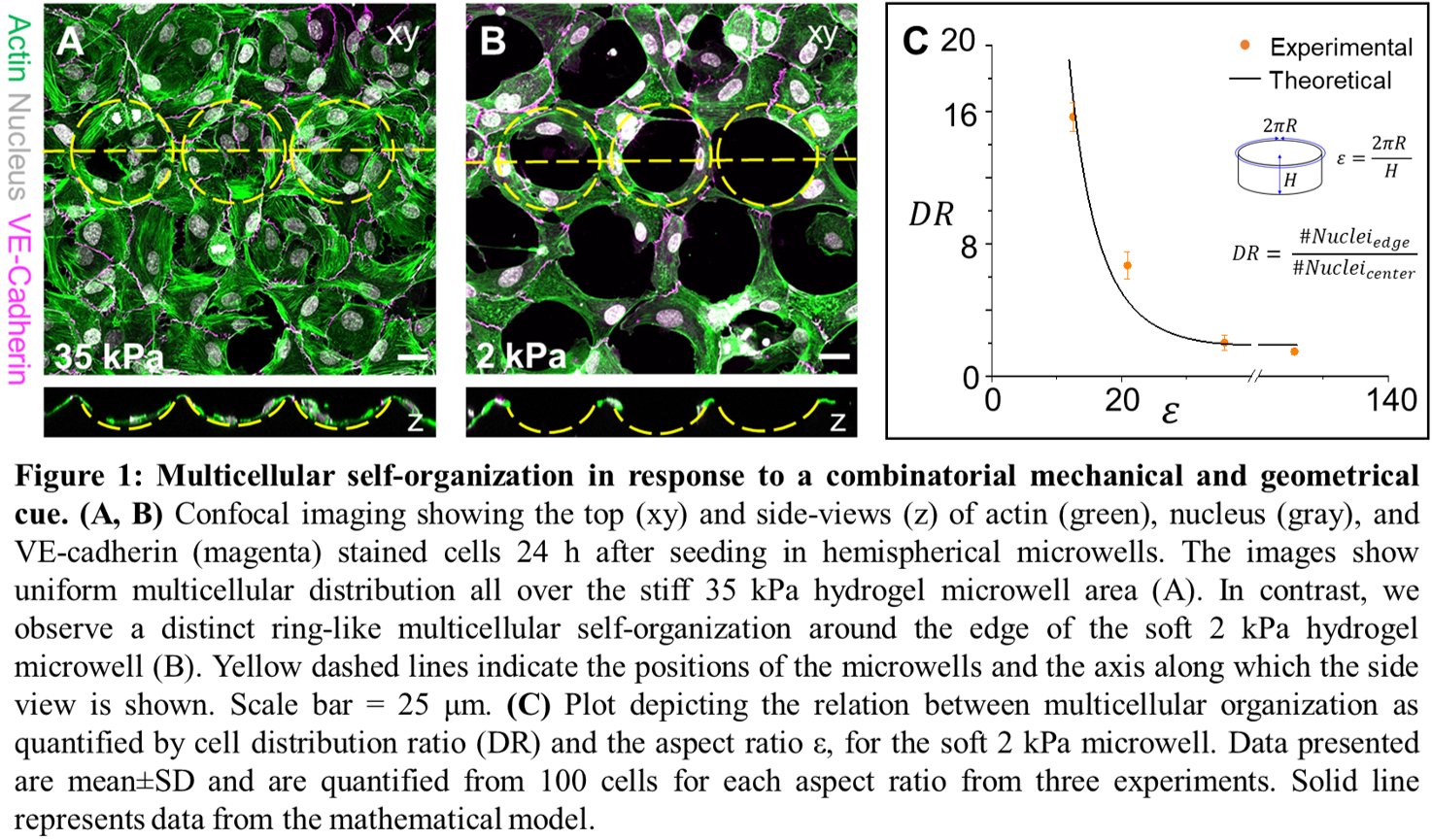
To investigate the effect of combinatorial cues, we microfabricated geometrically and mechanically tunable 3D microwells in gelatin hydrogels using photo- and soft lithography. We used scanning electron microscopy, Brillouin microscopy, rheometry, and atomic force microscopy to characterize the uniformity of these micropatterned hydrogels. We utilized live-cell time-lapse microscopy and quantitative epifluorescence/confocal microscopy to examine the self-organization of cells seeded to the hydrogel microwells.
We discovered that endothelial cells in soft 2 kPa microwells are approximately 30 times more likely to migrate to the microwell edge to organize in ring-like patterns than in stiff 35 kPa microwells (Fig 1A and B). We systematically examined this striking observation by varying the geometric and mechanical properties of the substrate (or the cell). We observed that the self-organization is independent of curvature and cross-sectional shape. Instead, we observe a quantitatively striking dependence on the microwell aspect ratio (perimeter/depth) when cells are growing on soft 2 kPa microwells with ring-like self-organization significantly more pronounced in microwells with aspect ratio < 25 (Fig 1C). Further, pharmacologically decreasing cytoskeletal tension for cells in stiff 35 kPa microwells led to self-organization of cells on the edges of the stiff 35 kPa microwells, similar to that in soft 2 kPa microwells, indicating that cells with lower cytoskeletal tension are sensitive to geometry. Following a discussion of these observations, this presentation will explain the mechanism leading to the striking differences in multicellular organization in response to combinatorial physical cues and a mathematical model which reveals that the ring-like self-organization results from the balance between tangential cytoskeletal tension, cell-cell, and cell-substrate adhesion. Finally, the presentation will show the technological implications of this new mode of self-organization which can be used to program reproducible cell patterning in customized CAD-designed microwell shapes, such as the letters of the word “CELL†simply by providing appropriate geometric and mechanical cues.
Our findings highlight the importance of the combinatorial effects of geometry and stiffness in complex cellular self-organization and provide a new method to guide the patterning of endothelial cells. We anticipate that this discovery will further facilitate the investigation of environmental cues on cell behavior and can be leveraged to design microenvironments that direct multicellular organization for complex devices, mimetic tissues, and organoid designs in the absence of protein patterning.