(631a) A Novel Microfluidic Device to Decipher the Interplay of Cellular Contractility and Electric Field during Galvanotaxis
AIChE Annual Meeting
2022
2022 Annual Meeting
Food, Pharmaceutical & Bioengineering Division
Cells, Organs, and Labs on a Chip
Thursday, November 17, 2022 - 12:30pm to 12:48pm
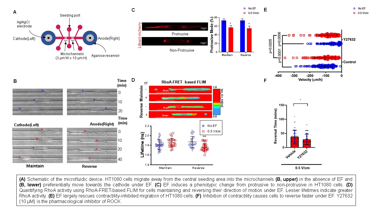
Materials and Methods: A polydimethylsiloxane (PDMS)-based microfluidic device was designed and fabricated using photolithography techniques and replica molding to create microchannels mimicking the narrow capillaries that the cancer cells travel through during metastasis. This was achieved by seeding cells in a central chamber of 3 µm width and 10 µm height (Fig. A), whereby the cells moved freely away from the reservoir into the microchannels. Ag/AgCl electrodes embedded in agarose were used at both ends of the device to apply DC electric fields across the microfluidic device using a programmable potentiostat. HT1080 fibrosarcoma cells were used in this study. Cell migration was visualized and recorded with time-lapse phase-contrast microscopy. The activity of proteins of interest was observed by using confocal microscopy along with fluorescently tagged proteins, such as FLIM-FRET biosensors or LifeAct tagged with mCherry. Pharmacological inhibitors were used to decipher migration mechanisms.
Results and Discussion: In the absence of an electric field, HT1080 fibrosarcoma cells migrated freely away from the central reservoir into the microchannels (Fig. B, upper). In the presence of electric field, we observe that the HT1080 cells, which were originally migrating towards the anode reversed their direction of migration after the EF was switched on, whereas the cells migrating towards the cathode maintained their direction of motion (Fig. B, lower). HT1080 cells subjected to EF undergo a phenotypic transition from a predominantly protrusive phenotype to a non-protrusive phenotype characterized by blunt, dense bundle of actin and blebs at the poles (Fig. C) in both the maintaining and reversing side of the device.
Since such phenotypic change is influenced by RhoA-ROCK-actomyosin-based contractility, we quantified RhoA activity in cells migrating in the maintaining and reversing side using the Förster resonance energy transfer (FRET)–based RhoA activity biosensor combined with confocal fluorescence lifetime imaging (FLIM). We found that the cells undergoing repolarization under EF have more RhoA activity unlike the cells on the maintaining side. This signified a difference in the role of RhoA-based contractility during the maintenance and reversal of migration direction under EF (Fig. D). On the maintaining side of the device, we found that EF facilitates the ROCK-inhibited migration of fibrosarcoma cells (Fig. E), in other words, EF bypasses the role of actomyosin-based contractility while maintaining the direction of cell migration under EF. However, on the reversing side, the inhibition of RhoA facilitates cell reversal, that is, contractility is acting as a hurdle for the EF to repolarize the cell (Fig. F).
Conclusion: We have designed and fabricated a novel microfluidic device that enables us to study the migration and reversal of directionality of cells in the presence of a direct-current electric field under physiologically relevant confinement. Using this device, we have uncovered the differences in the role that actomyosin-based contractility plays in maintaining and reversing the directionality of cells under EF. Our research will give more perspective into the different biophysical pathways that cells employ during galvanotaxis.
References:
[1] Huttenlocher, A., & Horwitz, A. R. (2007). Wound healing with electric potential. New England Journal of Medicine, 356(3), 303-304.
[2] Zhao, M. (2009, August). Electrical fields in wound healing—an overriding signal that directs cell migration. In Seminars in cell & developmental biology (Vol. 20, No. 6, pp. 674-682). Academic Press.
[3] Zhu, K., Hum, N. R., Reid, B., Sun, Q., Loots, G. G., & Zhao, M. (2020). Electric fields at breast cancer and cancer cell collective galvanotaxis. Scientific reports, 10(1), 1-11.
[4] Paul, C. D., Mistriotis, P., & Konstantopoulos, K. (2017). Cancer cell motility: lessons from migration in confined spaces. Nature Reviews Cancer, 17(2), 131-140.