(89c) Barrier-Free Paper Analytical Devices for Multiplex Colorimetric Detection
AIChE Annual Meeting
2022
2022 Annual Meeting
Topical Conference: Chemical Engineers in Medicine
Medical Devices
Monday, November 14, 2022 - 8:42am to 9:03am
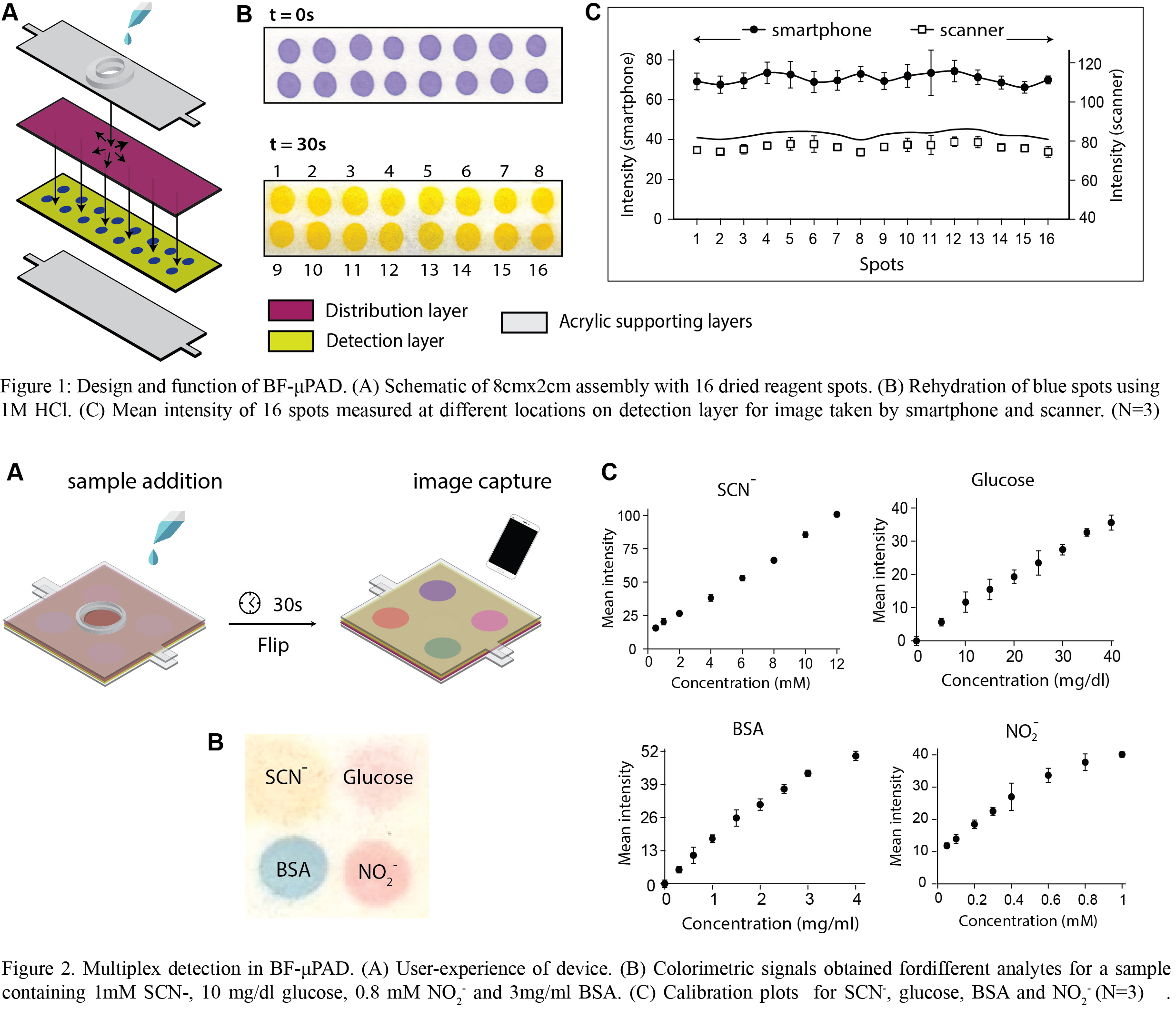
BF-μPADs consists of a stack of two paper layers of different wicking rates. The top layer is composed of Standard 17 glass fiber (called distribution layer) and offers less resistance to fluid flow. The bottom layer is composed of Whatman filter paper Grade 1 (called detection layer) and offers high resistance to fluid flow. The assembly was secured using acrylic supporting layers. Test reagents were deposited on the detection layer, and samples containing different analytes were added through the distribution layer. After the signals were fully developed, the device was flipped, and the colored signals were visualized using a smartphone in a custom-made imaging box and using a flatbed scanner. The image was then split into RGB color channels, and intensities of each spot were measured in the suitable channel using ImageJ software.
Prior to multiplex detection, a preliminary study was performed to investigate the uniformity of signals on the detection layer. 16 spots of bromophenol blue, a pH indicator dye, were deposited on an 8cmX2cm detection layer (Fig. 1A). An equal dimension of Standard 17 was placed flush on the detection layer, and 895ul (volumetric capacity of distribution and detection layer) of 1M HCl was added as the rehydrating fluid through the distribution layer. On addition of the acid, the pH of the dye changes, and the blue spots turn yellow. Figure 1B shows the colorimetric signals developed in the detection layer. It was observed that the intensities of the yellow color were similar and independent of the location of spots from the fluid source (Fig. 1C). The coefficient of variations of 48 spots across three device replicates was 6.94% for the smartphone and 3.49% for the scanner.
The multiplexing feature of BF-μPAD was demonstrated by testing salivary thiocyanate, glucose, nitrite, and bovine serum albumin (BSA) in a 2cm x 2cm assembly. Figure 2A shows the user experience of the device. After saturation of signals, the device was flipped, and the image of the detection layer was acquired using a smartphone. Figure 2B shows the colorimetric signals obtained in each spot for a sample containing 1 mM SCN-, 10 mg/dl glucose, 0.8 mM NO2- and 3 mg/ml BSA. Eight sample fluids with different concentrations of the four analytes were added to BF-μPAD, and mean test spot intensities (N=3 devices) were plotted against analyte concentrations. Intensities of all analytes increased monotonously with concentrations. Figure 2C shows the calibration plots of the four analytes.
We also compared the limit of detection (LOD) of the BSA assay performed in BF-μPAD and a traditional single-layer μPAD using a statistical method described by Holstein et al. (Holstein et al., 2015). LOD and the corresponding 95% confidence interval obtained using BF-μPAD were 0.1817 mg/ml and 0.1283-0.3682 mg/ml, respectively. The corresponding figures for μPAD were 0.6727 mg/ml and 0.5314-0.8217 mg/ml, respectively, signifying that BF-μPAD improved the LOD of the colorimetric assays by 3.7x compared to conventional μPAD. Further, a comprehensive study was performed to investigate the cross mixing of rehydrated signals on a longer assembly by depositing additional reagents upstream of the test signals. It was observed that the rehydrated signals from one chemistry do not mix with the downstream signals.
Barrier-free colorimetric detection in BF-μPAD is achieved by the unique flow pattern. The distribution layer rapidly wicks fluid in the lateral direction and continuously supplies it to the bottom detection layer from the vertical direction. Vertical entry of fluid practically eliminates lateral fluid movement in the detection layer, obviating the need for patterning barriers and generating spatially uniform signals. These flow patterns were modeled in COMSOL Multiphysics using Richards equation but are not presented here for brevity. Although the application of BF-μPAD was shown for biological fluid, the device can also be adapted to detect different pollutants in the environment, contaminants in soil, and pesticides in food. Since the distribution layer has large pores, the assembly is well suited for testing viscous emulsions like milk and denser fluids like honey which are challenging to flow alone through the cellulosic membrane of conventional μPADs.
BF-μPADs avert the need to pattern paper and enable faster flow and generate spatially uniform signals, improving upon the LOD of the assay. In contrast to μPADs, large-scale manufacturing of BF-μPADs would only require a commercially available benchtop robotic dispensing system to handle microliter volume.
This work is adapted with permission from Chauhan Ayushi, Toley Bhushan J., Anal. Chem. 2021, 93, 25, 8954–8961. Copyright 2021 American Chemical Society.