(89g) A Low Cost, Compact Flow Cytometer
AIChE Annual Meeting
2022
2022 Annual Meeting
Topical Conference: Chemical Engineers in Medicine
Medical Devices
Monday, November 14, 2022 - 10:06am to 10:27am
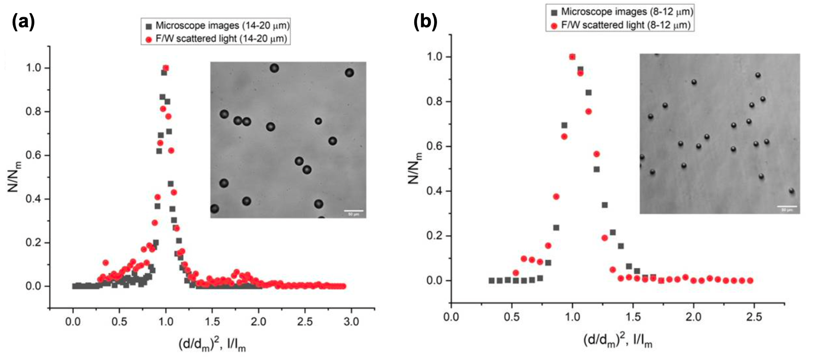
One of the critical parts of the cytometer is the flow cell where the particle stream is constrained into a tight region using hydrodynamic focusing. The conventional flow cells use thick quartz flow cells, they are expensive and therefore not suitable for instruments targeted for resource-constrained settings. We propose a low-cost sheath flow assembly design made of micro-tight ETFE adapters and an assembly of capillary tubes for the passage of focussed flow of particles. The entire flow cell assembly is a simplified version of a sheath flow cuvette design that is able to focus and accelerate a stream of particles in a narrow region probed by the incident laser light. The flow cell is embedded in a 3D-printed module, which houses microlenses and photodetectors, resulting in a compact flow cytometer. We show excellent agreement between the size distribution of model particles obtained via direct imaging and those obtained from light scattering. We have been able to obtain a 3-part differential of WBC based on the forward scatter measurement on our device. With minor modifications, the device may be used for disease detection, such as targeting viruses and bacteria.
The attached figure compares the size distribution of two different sets of particles obtained from image analysis of microscope images and that obtained from measurement of forward scattering data. The figure shows, (a) an overlap between the normalized Gaussian distribution of the cross-sectional area of 14-20ðœ‡m particles, (ð‘‘∕ð‘‘ð‘š)2, and the normalized Gaussian forward scattered intensity distribution, ð¼âˆ•ð¼ð‘š. Here, ð‘‘ð‘š is the mean value of the diameter of the particles in the mixture while ð¼ð‘š is the mean intensity value in the scattered light intensity distribution. The forward scatter intensity follows a direct square relationship with the particle diameter. A microscope image of the 14-20ðœ‡m particles in suspension has been provided in the inset for reference, (b): An overlap between the normalized Gaussian distribution of the cross-sectional area of 8-12ðœ‡m particles, (ð‘‘∕ð‘‘ð‘š)2 and the normalized Gaussian distribution of the forward scattered intensity, ð¼âˆ•ð¼ð‘š. A microscope image of the 8-12ðœ‡m particles in suspension has been provided in the inset for reference.