2023 AIChE Annual Meeting
(247b) Mussel-Inspired Mineralization of Centrifugally Spun 3D Fibers to Form a Tissue-Engineered Scaffold
Authors
The microstructure and the surface chemistry of the 3D biomineralized fibers were characterized by scanning electron microscopy (SEM) and attenuated total reflectance Fourier transform infrared spectroscopy (ATR-FTIR). The mechanical properties of the scaffolds were evaluated by compressing the samples in a wet state.
The biocompatibility of scaffolds was evaluated by in vitro cytotoxicity tests using an MTT assay on human mesenchymal stem cells (MSCs) cultured in the samplesâ extracts for 7 days. The Metabolism and viability of MSCs seeded on scaffolds were evaluated according to MTT analysis for up to 14 days. The morphology of MSCs cultured on the scaffolds was evaluated by scanning electron microscopy (SEM) 7 and 14 days after seeding.
The MSCs infiltration into scaffolds was evaluated by histological analysis assessments. Cell distribution within the scaffolds 7 and 14 days after cell seeding was evaluated using Hematoxylin and eosin (H&E) and DAPI staining and observed under an optical microscope.
The influence of the nanoâhydroxyapatite-based coating on gene expression of MSCs cultured on scaffolds after 14 days was evaluated using representative osteogenic markers.
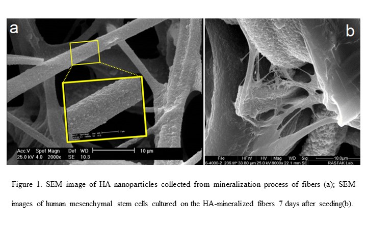
The results indicated uniform fibers with a smooth structure and represented the uniform formation of a polydopamine (PDA) ad-layer on the gelatin fibers. The thermally cross-linked fibers retained their structure and showed good dimensional stability under a wet state. SEM micrographs revealed that the nano-hydroxyapatite was formed uniformly over the nanofibers. The mineralization process enhanced the mechanical properties of scaffolds. All scaffolds exhibited high porosity (over 90%) and appropriate water uptake (approximately 1500%). All scaffolds exhibited appropriate biocompatibility, cell proliferation, migration, and infiltration. Mainly, the HA-mineralized fibrous scaffolds showed a favorable effect on the proliferation and differentiation of MSCs.The results suggest that a 3D porous mineralized fibrous scaffold could be a potential candidate for bone regeneration.