
-

-
Topics:
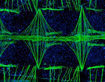
A fluorescent image of a mosquito's heart, taken by researcher Jonas King, took first place in the 35th annual Nikon's Small World contest.
This year scientists from 63 countries sent 2,200 entries into the competition. The submissions were judged not merely for their informational impact and technical proficiency but for their visual beauty. The judges, which included scientists and journalists, slowly whittled the entries down to the winner.
Jonas King, a member of the Vanderbilt research group studying the circulatory system of the Anopheles gambiae malarial mosquito, took the image (magnified 100x) that shows a section of it's tube-like heart.
Before he could take the winning picture, there was painstaking preparation. King explained the process to MSNBC's Cosmic Blog:
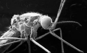
Preparing and photographing a mosquito's heart is an incredibly exacting job.
A slit has to be cut into the bug's abdomen. Its stomach and other organs have to be removed...King said it takes skill to carve up the mosquito for study. "I don't know if it's from playing guitar for all these years, but I'm good with my hands," he said.
Using a technique known as fluorescence microscopy, King used two types of fluorescent dyes, the green binds with the muscle cells and the blue with cellular DNA. By carefully tailoring the radiation shown on the speciman, he created a photograph with great scientific merit that shows the delicate traceries inside the heart. "This image of the heart helps us understand how they transport nutrients, hormones and even pathogens such as malaria throughout their bodies," said King.
According to Nikon, King's image was judged the winner for its combination of aesthetic beauty, scientific relevance and the technical difficulty in capturing it.
The full Vanderbilt press release here.
Nikon's official press release here.
Some of the other finalists included images of Zebrafish, a wasp's nest, a bird of paradise seed, red seaweed, Oacoxenite and more:
2rd Place, 2010:
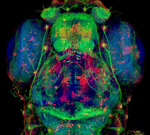
Dr. Hideo Otsuna of The University of Utah Medical Center, Dept. of Neurobiology and Anatomy, Salt Lake City, Utah, USA: 5-day old zebrafish head (20x) Confocal microscopy
Go to the Nikon Small World website to see the rest of the finalists...
Nikon MicroscopyU- source for information about various microscopy techniques
Comments
I just added to favorites your site. Good read.
Thanks. Glad you enjoyed the site.
I have to thank you for posting this, I like to hear about any good news.