
Technical Program for the 8th Bioengineering and Translational Medicine Conference
All times listed in PDT.
Details on the Poster Presentations can also be found here: https://www.aiche.org/BTM24/posters
Please note this schedule is subject to change. Check back for updates.
| Day 1: October 27, 2024 (Sunday) | ||
|---|---|---|
| 8:00 AM | 6:00 PM | Badge Pick-Up & On-site Registration |
| 8:00 AM | 8:30 AM | Morning Coffee & Networking |
| 8:30 AM | 8:40 AM | Welcome Remarks by the Conference Chairs: Jianping Fu, University of Michigan and Liangfang Zhang, UC San Diego |
| 8:40 AM | 10:30 AM | Session 1: Challenges & Opportunities in Drug Delivery Sesson Chair: Liangfang Zhang, UC San Diego |
| 8:40 AM | 9:10 AM | Keynote Speaker: Nicholas A. Peppas, The University of Texas at Austin "Reexamining Challenges & Opportunities in Drug Delivery, 1971 to now and beyond" Engineering the molecular design of intelligent biomaterials by controlling structure, recognition and specificity is the first step in coordinating and duplicating complex biological and physiological processes. Recent developments in siRNA and protein delivery have been directed towards the preparation of targeted formulations for protein delivery to specific sites, use of environmentally-responsive polymers to achieve pH- or temperature-triggered delivery, usually in modulated mode, and improvement of the behavior of their mucoadhesive behavior and cell recognition. We address design and synthesis characteristics of novel crosslinked networks capable of protein release as well as artificial molecular structures capable of specific molecular recognition of biological molecules. Molecular imprinting and micro-imprinting techniques, which create stereo-specific three-dimensional binding cavities based on a biological compound of interest, can lead to preparation of biomimetic materials for intelligent drug delivery, drug targeting, and tissue engineering. |
| 9:10 AM | 9:30 AM | Invited Speaker: Honggang Cui, Johns Hopkins University "From Prodrugs to Self-Assembling Prodrugs" Abstract: Covalent modification of therapeutic agents has historically been an effective method for enhancing a drug’s pharmacokinetic profile, leading to improved clinical outcomes. Integrating the concept of molecular assembly into prodrug design introduces a new dimension for the tailored synthesis of supramolecular nanomedicine to address unmet clinical needs. This self-assembling prodrug strategy uniquely and specifically exploits the self-assembling potential of therapeutic agents and the versatility in peptide design to create self-formulating and self-delivering prodrugs and prodrug hydrogelators, eliminating the need for excipients and additional carriers. In this presentation, I will detail our rational design of monodisperse, amphiphilic anticancer drugs that spontaneously assemble into discrete, stable supramolecular nanostructures with 100% prodrug loading. In some designs, the resultant nanostructures can be triggered to undergo a sol-gel transition in the biological environment. Our findings suggest that the formation of these nanostructures provides protection for both the drug and the biodegradable linker from the external environment, while also offering a mechanism for controlled release. Additionally, drug-based supramolecular hydrogels have been employed for the local delivery and sustained release of immune-modulating agents. This localized chemoimmunotherapy hydrogel demonstrates considerable potential to enhance antitumor immune responses and sensitize tumors to immunotherapies, providing a safer and more effective treatment approach. |
| 9:30 AM | 9:50 AM | Invited Speaker: Yoon Yeo, Purdue University "It’s still the delivery: Leveraging chemoimmunotherapy of cancer by bioactive ensembles" Abstract: Cancer immunotherapy exploits the immune system to control tumor growth, induce regression of established tumors, and develop antitumor immune memory to protect patients from recurrent diseases. Toward this goal, we have developed a nanoparticulate IMmunoActive compleX (called “IMAX”), comprising paclitaxel (PTX), an inducer of immunogenic cell death (ICD); 2E’, a carrier polymer with TLR5 agonist activity; and cyclic dinucleotide (CDN), a stimulator of interferon genes protein (STING) agonist. IMAX induces tumor-selective ICD, activates antitumor immune responses in various tumor models, and achieves tumor-free survival in 80-100% of the mice after a single intratumoral (IT) injection. Additionally, we have developed a poly(lactic-co-glycolic acid) (PLGA) NPs, decorated with adenosine triphosphate (ATP) and loaded with PTX for systemic administration, which recruits dendritic cells to the tumor and induces ICD in tumor cells, respectively. The PTX-loaded ATP-NP (PTX@ATP-NP) helped populate antitumor immune cells in the tumor microenvironment and attenuated tumor growth better than a mixture of PTX-loaded NP and ATP. However, these immunostimulatory treatments often face negative feedback from the host immune system, resulting in aggressive relapse in the long run. This presentation will demonstrate the rationale, development, and performance of different bioactive ensembles designed for chemoimmunotherapy of cancer and showcase some of our efforts to overcome evolving resistance. |
| 9:50 AM | 10:10 AM | Invited Speaker: Yichun Wang, University of Notre Dame "Engineering Biomimetic Materials to Empower Therapeutic Exosomes as Future Drug Delivery Platform" Exosomes are a subset of extracellular vesicles, with diameters between 50 nm and 150 nm, secreted by most eukaryotic cells. They are promising drug delivery vehicles due to their small size, biocompatibility, low immunogenicity, and reduced toxicity in comparison with synthetic nanoscale formulations such as liposomes, dendrimers, and polymers. However, there remain fundamental challenges to the utilization of exosomes in the clinic: i) drug loading efficiency into exosomes is very limited; ii) the production of exosomes has yet to reach sufficient high throughput for clinical tests or even further development; iii) endowing exosomes with multiple abilities for satisfactory disease targeting, tracking and combinational therapies is highly demanding. In this talk, I will introduce a convergent bioengineered platform enabled by engineered biomimetic materials developed in The Wang Lab at the University of Notre Dame for advancing therapeutic exosomes in future medicine. This platform includes 1) A high-efficiency exosome drug loading technology with chiral graphene nanoparticles; 2) A high-yield in vitro exosome production cell culture scaffold with stimulating piezoelectric nanofibers; 3) Engineered hybrid exosomes with biomimetic nanoparticles as a multifunctional targeted delivery system for cancer treatment. The platform allows loading drugs into exosomes with high efficiency, biomanufacturing exosomes in high throughput, and further engineering exosome-based drug delivery systems for various diseases with desired functions including targeted delivery, imaging, and multifunctional therapies. |
| 10:10 AM | 10:20 AM | Submitted Abstract: Nicole Henry, Imperial College London "Leveraging a Clot Component As the Starting Material for Targeted Delivery of Tissue Plasminogen Activator to Treat Cardiovascular Diseases" Abstract: Designing a specific and targeted drug delivery system (DDS) is crucial for its success. By leveraging one of the main components of blood clots, red blood cells, to produce naturally derived nanoparticles (RBCVs), a level of complexity, unattainable by synthetic materials, can be achieved. RBCVs will inherit properties from their source: long circulation, biocompatibility and biodegradability. Cardiovascular diseases are the leading cause of death. Main cardiovascular disorders, including strokes, are initiated by thrombus formation. Currently, there is only one FDA approved treatment for acute ischemic stroke; tissue plasminogen activator (tPA), which has several limitations: short half-life, unwanted bleeding complications, <50% recanalization rate for patients those who receive treatment and short treatment window. By loading tPA into RBCVs and functionalizing the surface with a targeting peptide, cyclic arginyl glycyl-L-aspartic acid (cRGD) (cRGD-tPA-RBCVs), a clot specific thrombolytic nanoparticle was produced overcoming the abovementioned limitations of the free therapeutic. cRGD was chosen as the targeting ligand for this system as it is known to preferentially bind to the GPIIb-IIIa integrin present on activated platelets. By using a component of the clot as a raw material, a novel homing effect was observed, resulting in a bi-specific clot targeting DDS. cRGD-tPA-RBCVs have demonstrated specific binding to activated platelets (>85%), as well as thrombolysis in numerous in vitro assays. This system has been further validated with in vivo models. It has shown good pharmacokinetics (t1/2 > 50 minutes) and biodistribution as well as enabling thrombolysis and enhancing clot penetration (>4-fold increase), leading to successful blood flow recovery (>75%). These results indicate the promise of a clot specific natural nanoparticle for stroke treatment by selectively delivering tPA to thrombi through a targeting ligand and novel homing effect. |
| 10:20 AM | 10:30 AM | Submitted Abstract: Jiahe Li, University of Michigan "Nonpathogenic E. coli Engineered to Surface Display Cytokines As a New Platform for Immunotherapy" Abstract: Given the safety, tumor tropism, and ease of genetic manipulation in non-pathogenic Escherichia coli (E. coli), we designed a novel approach to deliver biologics to overcome poor trafficking and exhaustion of immune cells in the tumor microenvironment, via the surface display of key immune-activating cytokines on the outer membrane of E. coli K-12 DH5α. Bacteria expressing murine decoy-resistant IL18 mutein (DR18) induced robust CD8+ T and NK cell-dependent immune responses leading to dramatic tumor control, extending survival, and curing a significant proportion of immune-competent mice with colorectal carcinoma and melanoma. The engineered bacteria demonstrated tumor tropism, while the abscopal and recall responses suggested epitope spreading and induction of immunologic memory. E. coli K-12 DH5α engineered to display human DR18 potently activated mesothelin-targeting CAR NK cells and safely enhanced their trafficking into the tumors, leading to improved control and survival in xenograft mice bearing mesothelioma tumor cells, otherwise resistant to NK cells. Gene expression analysis of the bacteria-primed CAR NK cells showed enhanced TNFα signaling via NFkB and upregulation of multiple activation markers. Our novel live bacteria-based immunotherapeutic platform safely and effectively induces potent anti-tumor responses in otherwise hard-to-treat solid tumors, motivating further evaluation of this approach in the clinic. |
| 10:30 AM | 11:00 AM | Break Sponsored by the journal Bioengineering & Translational Medicine (BioTM) supported by Wiley |
| 11:00 AM | 12:50 PM | Session 2: Biomanufacturing Session Chair: Y. Shrike Zhang, Harvard Medical School |
| 11:00 AM | 11:30 AM | Keynote Speaker: Sarah Heilshorn, Stanford University "Designer biomaterials to enable diffusion-based biofabrication" Abstract: The controlled use of diffusion is an emerging strategy to tailor the properties and geometry of bioprinted constructs. During embedded bioprinting, molecules with specialized functions, including crosslinkers, catalysts, and viscosity-modifiers, can diffuse across the interface between the printed construct and the surrounding support bath. This molecular diffusion can be designed to modify the construct material properties, including the microstructure, stiffness, and biochemistry, all of which impact cell phenotype. Thus, this new category of diffusion-based biofabrication opens up new opportunities to guide biological functionality. Here, I present several recent biomaterials my group has designed for use as bioinks in these new diffusion-based strategies. In one example, we modified four different types of biopolymers to display reactive pendant groups that can participate in bio-orthogonal click reactions. Upon diffusion of crosslinkers with a compatible bio-orthogonal reactive group, a single construct composed of multiple bioinks is readily formed. Importantly, the interfaces within these multi-material constructs are uniform, cohesive, and strong. In a second example, we use radial diffusion across an interface to create perfusable structures with complex branching patterns. By controlling the diffusion time, we can fine-tune both the inner and outer diameters of each segment within the perfusable network. These tissue models are being used to mimic vascular-like and fallopian-tube like structures. Finally, I present a family of bioinks crosslinked with dynamic covalent bonds. These viscoelastic biomaterials have mechanical properties that mimic the extracellular matrix; however, these materials are quite challenging to use as bioinks due to their unique rheological properties. To solve this challenge, we use the temporary presence of catalysts and crosslink inhibitors to reversibly modify the scaffold mechanical properties. These scaffolds are being used to mimic the fibrosis and matrix stiffening that occurs during liver disease and cancer progression. Taken together, these examples demonstrate how diffusion-based bioinks can be harnessed in the biofabrication of tissues with biomimetic functionality. |
| 11:30 AM | 11:50 AM | Invited Speaker: Sean Palecek, University of Wisconsin-Madison "Predicting Outcomes and Improving Reliability of Manufacturing Cardiomyocytes Based on Multi-Omic Characterization Of Pluripotent Stem Cell-Derived Cardiac Progenitors" Abstract: Human pluripotent stem cells (hPSCs) provide a limitless source of human cell and tissues for modeling development and disease, evaluating the safety and efficacy of molecular therapeutics, and for developing cell therapies. For example, hPSC-derived cardiomyocytes are used in the drug development pipeline to identify cardiotoxicity and efficacy in a human model and are in clinical trials to restore cardiac function in patients with heart failure. Challenges associated with cardiomyocyte manufacturing, including difficulties in scaling processes to those needed for commercial and clinical applications and significant batch-to-batch variability, limit the application of these cells however. hPSCs transition through multiple progenitor states during cardiomyocyte differentiation. One of these states, termed the cardiac progenitor cell (CPC), is a multipotent cell type committed to the cardiac fate but capable of generating multiple cell types of the heart including cardiomyocytes, cardiac fibroblasts, smooth muscle cells, and epicardial cells. We identified CPC populations that give rise to high percentages of cardiomyocytes (high efficiency CPCs) and CPC populations that generate low purity cardiomyocytes (low efficiency CPCs), then performed an integrated transcriptomic (RNAseq) and epigenomic (ATACseq) analysis of these CPCs to identify features that predict CM generation. By comparing our datasets to those in the literature, we found a set of genes enriched in high efficiency CPCs.Our comparison of pathway enrichment in high and low efficiency CPCs also suggested MAP kinase and WNT signaling as significant drivers of off-target cell population generation, including skeletal myocytes, epicardial cells and a SLC7A11-expressing population of unknown cells. We found that stage-specific modulation of these pathways improves reliability of CM manufacturing. In conclusion, our study demonstrates how integrated multi-omics analysis of progenitor cells can identify quality attributes that enable prediction of cell differentiation potential, thereby improving differentiation protocols and increasing manufacturing robustness of cell therapies. |
| 11:50 AM | 12:10 PM | Invited Speaker: Shaochen Chen, University of California, San Diego "Rapid 3D Bioprinting for Precision Tissue Models" Abstract: In this talk, I will present our laboratory’s recent research efforts in developing rapid, digital light processing (DLP) based bioprinting methods to create 3D tissue constructs using a variety of biomaterials and cells. These 3D printed scaffolds are functionalized with precise control of micro-architecture, mechanical (e.g. stiffness), chemical, and biological properties. Such functional tissues allow us to investigate microscale cell-microenvironment interactions in response to integrated mechanical and chemical stimuli. From these fundamental studies we have been creating both in vitro and in vivo precision tissues for tissue regeneration, disease modeling, and drug discovery. Examples including 3D bioprinted liver and heart models will be discussed. I will also showcase 3D printed biomimetic scaffolds for nerve repair. Throughout the presentation, I will discuss engineer’s perspectives in terms of design innovation, biomaterials, mechanics, and scalable biomanufacturing. 1. You et al, “High Cell Density and High Resolution 3D Bioprinting for Fabricating Vascularized Tissues”, Science Advances, 9 (no. 8), eade7923, 2023. 2. Koffler et al., “Biomimetic 3D-Printed Scaffolds for Spinal Cord Injury”, Nature Medicine, 25, 263-269, 2019 3. Ma et al., Deterministically Patterned Biomimetic Human iPSC-derived Hepatic Model via Rapid 3D Bioprinting”, PNAS, 113 (no. 8), pp. 2206-2211, 2016. |
| 12:10 PM | 12:30 PM | Invited Speaker: Y. Shrike Zhang, Harvard Medical School "Unconventional Additive (Bio)Manufacturing Methods for Regenerative Medicine" Abstract: Over the last decades, the field of three-dimensional (3D) printing, or additive manufacturing, has witnessed tremendous progress. 3D printing enables precise control over the composition, spatial distribution, and architecture of the printed constructs facilitating the recapitulation of the delicate shapes and structures of target patterns. More recently it has been further combined with cells and cell-laden biomaterials to offer the versatility to fabricate biomimetic volumetric tissues that are both structurally and functionally relevant. Nevertheless, conventional 3D printing and bioprinting techniques are limited in certain aspects. This talk will thus discuss our recent efforts in developing a series of advanced additive (bio)manufacturing strategies that take unconventional approaches to tackle some of these problems and improve their capacities towards diverse applications in biomedicine with a focus on regenerative medicine. These platform technologies will likely provide new opportunities in areas from constructing functional tissues and microtissue models for promoting personalizable medicine, all the way to minimally invasive surgical implications. |
| 12:30 PM | 12:40 PM | Submitted Abstract: Hung Pang Lee, Stanford University "Bioprinting of Microribbon-Shape Microgels with Tunable Stiffness for Stem Cell Differentiation and Hierarchical Patterning" Abstract: Bioprinting enables precise spatial placement of cells and biomaterials to create complex tissues. Microribbon (µRB)-shaped microgels, which our group previously reported, crosslink into macroporous hydrogels offering advantages like interconnective microporosity and high surface area for cell adhesion, migration, proliferation, and differentiation[1]. This study developed µRB bioinks with tunable stiffness for extrusion-based bioprinting and explored their applications in stem cell differentiation and hierarchical patterning for modeling cancer invasion and microvessel formation. Gelatin-based µRBs were fabricated using wet-spinning and crosslinked with glutaraldehyde. To tune µRB stiffness, three levels of amine group crosslinking were used: 5%, 10%, and 15%. Methacrylate-functionalized µRBs were photo-crosslinked into 3D scaffolds after bioprinting. The µRB bioinks and printed constructs were assessed for mechanical properties, printability, and biocompatibility. Embedded human mesenchymal stem cells (hMSCs) were evaluated for osteogenic differentiation and bone formation over 28 days. Hierarchical patterning of µRB bioink was applied to construct a 3D breast cancer-bone metastasis model, which we used to compare invasion behavior of two breast cancer cell lines. The effects of µRB stiffness on cell alignment in bioprinting were also evaluated. µRB bioinks with three stiffness levels showed favorable printability and supported high cell viability post-bioprinting. Increased µRB stiffness enhanced hMSC osteogenesis and bone formation. A spatially-patterned 3D breast cancer invasion model was successfully bioprinted and demonstrated differential invasion potential by two cell lines.. The anisotropic µRBs promoted cell alignment, which increased with higher stiffness due to the increased shear stress during extrusion. This alignment facilitated the formation of microvessel-like structures when bioprinting hMSCs and endothelial cells. In conclusion, µRB bioinks with tunable stiffness support robust stem cell differentiation and complex 3D tissue hierarchical construction for modeling cancer invasion and microvessel formation. [1] Han, Li‐Hsin, et al. Advanced Functional Materials 23.3 (2013): 346-358. |
| 12:40 PM | 12:50 PM | Submitted Abstract: Saskia Fogg, University of Birmingham, Bayer Pharmaceuticals "Optimising Embedded Bioprinting Support Matrix Formulation to Improve the Resolution of 3D-Printed Skin Implants" Abstract: Suspended layer additive manufacturing (SLAM) is an embedded bioprinting technique that utilises fluid gels as a temporary support matrix to facilitate the extrusion printing of soft hydrogel-based bioinks into complex, functional, three-dimensional (3D) tissue structures. Here, we report the use of SLAM to produce a continuous tri-layered skin implant which closely resembles healthy human skin. Composed of micrometre-sized gel particles, fluid gels behave in bulk as viscoelastic fluids with shear-thinning and self-healing properties. This enables the support matrix to act as a liquid as the cartridge needle passes through it, yet rapidly recover solid-like properties to support the deposited bioink, preventing the spread and collapse of the printed structure until it can be stabilised. SLAM is a novel technique that expands the potential for bioprinting and tissue engineering but also complicates print parameter characterisation and optimisation by introducing additional parameters to an already complex process. The rheological and physical properties of both the support matrix and bioink are critical parameters that significantly impact the resolution and fidelity of the final construct. These properties can be modified and controlled by formulation and process parameters. Here, we aim to 1) characterise agarose fluid gel support matrix formulations and alginate and/or collagen bioink formulations through rheological, mechanical, and particle size analysis. 2) compare how these parameters correlate with the resolution of a printed construct through profilometry. 3) print a tri-layered skin implant with greater resolution than previous work. |
| 12:50 PM | 2:00 PM | Lunch |
| 2:00 PM | 3:30 PM | Session 3: Cell Therapy & Protein Engineering |
| 2:00 PM | 2:30 PM | Keynote Speaker: David Schaffer, University of California, Berkeley "Directed Evolution of New AAV Vectors for Clinical Gene Therapy" Abstract: Gene therapy has experienced an increasing number of successful human clinical trials, leading to 6 FDA approved products using delivery vectors based on adeno-associated viruses (AAV). These clear successes have been made possible by the identification of disease targets that are suitable for the delivery properties of natural variants of AAV. However, vectors in general face a number of barriers and challenges that preclude their extension to most disease targets, including pre-existing antibodies against AAVs, suboptimal biodistribution, limited spread within tissues, an inability to target delivery to specific cells, and/or limited delivery efficiency to target cells. These barriers are not surprising, since the parent viruses upon which vectors are based were not evolved by nature for our convenience to use as human therapeutics. Unfortunately, for most applications, there is insufficient mechanistic knowledge of underlying virus structure-function relationships to empower rational design improvements. As an alternative, for two decades we have been implementing directed evolution – the iterative genetic diversification of the viral genome and functional selection for desired properties – to engineer highly optimized, next generation AAV variants for delivery to any cell or tissue target. We have genetically diversified AAV using a broad range of approaches from fully random (e.g. error prone PCR) to computationally guided (e.g. by machine learning). The resulting large (~109) libraries are then functionally selected for substantially enhanced delivery, yielding AAVs capable of highly efficient therapeutic gene delivery in animal models and in several human clinical trials. |
| 2:30 PM | 2:50 PM | Invited Speaker: Xiaoping Bao, Purdue University "Engineer CAR-neutrophils for targeted chemoimmunotherapy against glioblastoma" Abstract: Glioblastoma (GBM), the most common type of primary brain tumor, is characterized by high mortality rate, short lifespan, and poor prognosis with a high tendency of recurrence. Functional therapeutics, including PRMT5 inhibitors, radiosensitizers, and emerging chimeric antigen receptor (CAR)-T immunotherapy, have been developed to treat GBM. However, the existence of physiological blood-brain barrier (BBB) or blood-brain-tumor barrier has impeded the efficient delivery of such promising therapeutics into the brain and limited their therapeutic efficacy. Given the native ability of neutrophils to cross BBB and penetrate the brain parenchyma, here we tested the therapeutic concept that neutrophils could be engineered with synthetic CARs to specifically target GBM and effectively deliver chemo-drugs to brain tumor as a novel dual chemoimmunotherapy for the first time. Primary neutrophils are short-lived and resistant to genetic modification. Therefore, we genetically engineered human pluripotent stem cells with different chlorotoxin (CLTX) CARs and differentiated them into functional CAR-neutrophils. As compared to CAR-natural killer (NK) cells, systemically administered hPSC-derived CLTX CAR-neutrophils significantly reduced tumor burden in xenograft mouse models and extended their lifespan, suggesting superior abilities of neutrophils in crossing BBB and penetrating GBM xenograft in mice. We also loaded hypoxia-activated prodrug tirapazamine (TPZ) into CAR-neutrophils using silica nanoparticles with rough surfaces (R-SiO2-TPZ) and demonstrated their enhanced antitumor activities in xenograft mouse models, serving as a novel dual chemoimmunotherapy against GBM. Our results established that CAR neutrophil-mediated drug delivery may provide an effective and universal strategy for specific targeting of solid tumors. |
| 2:50 PM | 3:10 PM | Invited Speaker: Juliane Nguyen, University of North Carolina "Sticky Solutions: Advancing Targeted Therapies" |
| 3:10 PM | 3:20 PM | Submitted Abstract: Qing Shao, University of Kentucky "S-Plm: Structure-Aware Protein Language Model Via Contrastive Learning between Sequence and Structure" Abstract: Proteins play an essential role in various biological and engineering processes. Large protein language models (PLMs) present excellent potential to reshape protein research by accelerating the determination of protein function and the design of proteins with the desired functions. The prediction and design capacity of PLMs relies on the representation gained from the protein sequences. However, the lack of crucial 3D structure information in most PLMs restricts the prediction capacity of PLMs in various applications, especially those heavily dependent on 3D structures. To address this issue, we introduce S-PLM, a 3D structure-aware PLM that utilizes multi-view contrastive learning to align the sequence and 3D structure of a protein in a coordinated latent space. Additionally, we provide a library of lightweight tuning tools to adapt S-PLM for diverse protein property prediction tasks. Our results demonstrate S-PLM’s superior performance over sequence-only PLMs on all protein clustering and classification tasks, achieving competitiveness comparable to state-of-the-art methods requiring both sequence and structure inputs. |
| 3:20 PM | 3:30 PM | Submitted Abstract: Nicholas Hutchins, MIT "Reconstructing Signaling History and Spatial Organization of Single Cells Via Generalizable Statistical Inference" Abstract: Proper cell-cell communication is the linchpin for the development of multicellular organisms. Mapping these communication networks is crucial for understanding the logic of embryonic development and for directing embryonic stem cells differentiating into desired fates. In the past, cell-cell communication has been primarily mapped through time-consuming animal genetics. Here, by fitting conditional variational autoencoders (CVAE) to scRNA-seq data, we created IRIS (Intracellular Response to Infer Signaling state), a semi-supervised deep learning method for annotating signaling state of individual cells only using its gene expression. Compared to other cell communication prediction algorithms based on ligand-receptor expression, IRIS relies on the induced gene expression changes in signal-receiving cells, which serves as the most accurate measure for functional cell-cell communication. However, such inference is complicated by the perceived context-dependent nature of gene expression changes. We demonstrate that many developmental signaling pathways induce cell type independent signatures of gene expression. This enables us to train our model on limited datasets and predict the signaling state of previously unseen cell types. The predictions are highly accurate, as quantified by orthogonal datasets we generated, in which human embryonic stem cells were differentiated upon sequential combinatorial ligand stimulation. We validated that the predictions recovered existing knowledge of cell communication in diverse biological contexts. In contrast to other signal prediction algorithms, IRIS requires no observation of ligand sending cells or hard labels on the clusters for prediction, allowing heterogeneous ligand stimulation to be accurately annotated at the single-cell level. Furthermore, we show that these predictions can be used to annotate signaling history and reconstruct the spatial relationship amongst cells. We anticipate that this tool will be generalizable to solve a wide class of cell-cell communication inference problems and provide better defined signaling conditions for stem cell differentiation. |
| 3:30 PM | 4:00 PM | CIRM Presentation: James Campanelli, California Institute for Regenerative Medicine |
| 4:00 PM | 4:30 PM | Break |
| 4:30 PM | 5:40 PM | Session 4: AI & Data Science Session Chair: Jianping Fu, University of Michigan |
| 4:30 PM | 5:00 PM | Keynote Speaker: Tara Deans, Georgia Tech "Harnessing Synthetic Biology for Next-generation Therapeutics" Abstract: Synthetic biology has transformed how cells can be reprogrammed, providing a means to reliably and predictably control cell behavior with the assembly of genetic parts into more complex synthetic gene circuits. Using these approaches, we are programming stem cells with novel genetic tools to control genes and pathways that result in changes in their native function for desired outcomes. We are particularly interested in building genetic tools to define the molecular rules governing hematopoietic cell fate transitions and the dynamic processes that guide stem cell differentiation. We have also shown that megakaryocytes, the progenitor cells for platelets, can be reprogrammed and loaded with non-native proteins to produce engineered platelets that function as local and systemic delivery devices. |
| 5:00 PM | 5:20 PM | Invited Speaker: Zeinab Jahed, University of California, San Diego "Intelligent Nano-electronics for Intracellular Sensing" Abstract: Electrical signaling through voltage gated ion channels is the quickest form of communication between cells in tissues like the brain, heart, and pancreas. This signaling appears as swift changes in the electrical potential inside cells. These signals are difficult to detect due to their rapid and small nature, usually requiring invasive techniques like patch clamping, which involve piercing the cell membrane to reach the interior. In this study, we introduce an innovative method that combines nanotechnology and physics-informed AI to detect intracellular electric potentials non-invasively, avoiding the need to penetrate the cell membrane, enabling stable term recordings. Our technology has broad applications in drug cardiotoxicity screening and electrophysiological research. |
| 5:20 PM | 5:40 PM | Invited Speaker: Adam Gromley, Rutgers University "Automation and Active Learning for the Autonomous Design of Polymer Biomaterials" Abstract: The seamless integration of synthetic materials with biological systems long remains a grand challenge, often curtailed by the sheer complexity of the cell-material interface. For decades, biomaterial scientists and engineers have designed around this complexity by rationally designing new materials one experiment at a time. However, recent advances in laboratory automation, high throughput analytics, and artificial intelligence / machine learning (AI/ML) now provide a unique opportunity to fully automate the design process. In this seminar, we put forth our efforts to develop a biomaterials acceleration platform (BioMAP) (i.e., self-driving biomaterials lab) that can rapidly iterate through design spaces and identify unique material properties that perfectly synergize with biological complexity. |
| 5:40 PM | 6:00 PM | Invited Speaker: Daniel Reker, Duke University "Machine Learning for Drug Discovery and Delivery" Abstract: Machine learning is increasingly applied in drug development to accelerate early discovery but faces challenges due to limited high-quality datasets for more complex development stages. We are developing yoked learning approaches and molecular pairwise representations to further enhance machine learning performance, particular for deep neural networks and complex ADMET endpoints. Similar data scarcity challenges are observed in drug delivery, where complex materials and small datasets have prohibited broader deployment of machine learning solutions. To combat these challenges, we are developing novel materials and machine learning platforms to create safer and more effective drug formulations. |
| 6:00 PM | 6:05 PM | Sponsored Talk: CorDx |
| 6:05 PM | 7:30 PM | Reception & Poster Session: Cocktail Reception sponsored by CorDx |
| Day 2: October 28, 2024 (MONDAY) | ||
|---|---|---|
| 8:00 AM | 12:00 PM | Badge Pick-Up & On-site Registration |
| 8:00 AM | 8:30 AM | Morning Coffee & Networking |
| 8:30 AM | 8:40 AM | Morning Remarks by the Conference Chairs: Jianping Fu, University of Michigan and Liangfang Zhang, UC San Diego |
| 8:40 AM | 10:30 AM | Session 5: Women's Health Session Chair: Zongmin Zhao, University of Illinois at Chicago |
| 8:40 AM | 9:10 AM | Keynote Speaker: Lonnie Shea, University of Michigan "Detection and Treatment of Undesired Immune Responses" Abstract: Chronic disease is now responsible for 7 of every 10 deaths in the United States, increasing due to the widespread availability of antibiotics, antivirals, and vaccines for treating infectious disease. Patients with infectious disease develop acute symptoms that can be monitored using devices, such as a thermometer in the case of fever. An increased temperature both informs therapeutic decision making and indicates that the immune system is taking healthy action against the pathogen. Unlike infectious diseases, chronic diseases (cancer, diabetes, Alzheimer’s, etc.) develop slowly and asymptomatically, and often go undetected until the function of a vital organ has been impacted. Central to many chronic diseases are undesired immune responses that either contribute to disease progression or limit the efficacy of therapies. |
| 9:10 AM | 9:30 AM | Invited Speaker: Mana Parast, University of California, San Diego "Stem Cell-based Modeling of the Human Placenta" Abstract: The human placenta is a transient organ, key for proper fetal growth and development. Abnormal development and function of this organ is associated with adverse pregnancy outcomes, including pre-eclampsia, fetal growth restriction, and stillbirth. The human placenta is also unique in its structure, cellular composition, and signaling pathways, particularly in the early embryonic period, thus limiting the utility of animal models for its study. Specification and early development of trophoblast, the epithelial cells of the placenta, have been the subject of intense study over the past decade, with the advent of single cell and spatial ‘omics technology and their convergence on the fields of human embryology, pluripotent stem cell, and placental biology. In particular, establishment of culture conditions for derivation of trophoblast stem cells (TSC) and trophoblast organoids (TOrg) from both placental tissues and pluripotent stem cells has significantly advanced our ability to study trophoblast differentiation. Nevertheless, many questions remain, including whether these models reflect trophoblast differentiation in vivo. In addition, significant barriers exist regarding access to these model systems and funding for such research. It is therefore most important to identify the strengths and limitations of each of these models, while simultaneously educating the public about this research and advocating for reasonable regulation and increased funding. Over the past decade, our group has optimized pluripotent stem cell-derived models for trophoblast differentiation, in particular, asking whether they recapitulate diseased trophoblast, while also comparing them to primary (placenta-derived) cells from placentas of different gestational ages. Overall, our data reveal significant differences between established TSC and the vast majority of primary cytotrophoblast progenitor cells in early gestation placenta. At the same time, we see a high level of similarity between primary and pluripotent stem cell-derived TSC, confirming the latter as a potentially robust, easily-accessible model for studying human trophoblast differentiation. |
| 9:30 AM | 9:50 AM | Invited Speaker: Brian Aguado, University of California, San Diego "Determining Sex Differences in Cardiovascular Diseases Using Biomaterials" Abstract: Cardiovascular disease is the leading cause of death in both men and women, yet our mechanistic knowledge of the sex-specific molecular and cellular mechanisms that guide cardiovascular disease progression, particularly in women, remain poorly characterized. Studies evaluating disease mechanisms rarely state the sex of cells used for in vitro studies or are performed primarily in male animal models, causing our gap in knowledge. My laboratory uses precision biomaterials as in vitro and in vivo tools to dissect mechanisms that contribute to sexual dimorphisms in cardiovascular diseases, specifically aortic valve stenosis. In my talk, I will discuss how we have used hydrogel biomaterials as engineered valve matrix mimics to explore sex dimorphisms in valvular interstitial cell phenotypes in vitro and describe sex-specific molecular mechanisms that may drive dimorphisms in aortic valve stenosis. Our work seeks to leverage biomaterial technologies to understand sex dimorphisms in health and disease, with the long-term goal of achieving sex and gender equity in cardiovascular disease treatments and outcomes. |
| 9:50 AM | 10:00 AM | Submitted Abstract: Emily Du, University of Washington "Nicotinamide-Loaded Nanopeptoids for Energy Regeneration to Drive DNA Repair in Neonatal Brain Injury" Abstract: Neonatal brain injury results in DNA damage, which activates poly(ADP-ribose) polymerase-1 (PARP-1) and decreases cytosolic nicotinamide adenine dinucleotide (NAD+), decreasing adenosine triphosphate (ATP) production, impairing mitochondrial function, and inactivating cellular metabolism, leading to cell death. Increasing NAD+ levels could therefore restore cellular NAD+ to aid in DNA repair, and increase cell survival. In the brain, NAD+ is more likely to be made by nicotinamide (NAM)-derived nicotinamide mononucleotide (NMN) available locally to brain cells. However, mammalian cells cannot import NAD+, and the cell-specific targeting of NAD+ and NAM is limited. Therefore, cellular delivery strategies are needed to capture the potential of an NAD+-restorative therapeutic approach. We have developed a nanopeptoid delivery strategy to replenish cellular redox state and energy production (Fig. 1A) following acute neonatal brain injury. Peptoids, a synthetic analog of peptides, are sequence-specific heteropolymers that were developed as protein mimetics possessing advantages of both synthetic polymers and biopolymers. We self-assemble peptoids into tubular form to create peptoid nanotubes (PNTs) of varying lengths (Fig. 1B) that can extravasate more readily and penetrate through the cell membrane. PNTs conjugated with NAD+ or NAM achieved high drug loading efficiency and no cytotoxicity in brain cells (Fig. 1C). NAM-PNTs localize in microglia and associate with neurons (Fig. 1D). NAM-PNTs improve cell viability in response to oxygen glucose deprivation (OGD) in organotypic brain slices (Fig. 1E). NAM-PNTs replenish intracellular ATP levels by 24h after treatment (Fig 1F) and drive glial proliferation (Fig 1G). We see an associated shift in reduced pro-inflammatory cytokine production and increase anti-inflammatory cytokines. In the postnatal day 10 (P10) hypoxic-ischemic rat, a single dose of NAM-PNTs administered systemically decreased brain tissue area loss and improved neuropathology. Our study demonstrates that NAM delivery via PNTs has strong therapeutic potential for cell-specific delivery and energy regeneration in the acutely injured neonatal brain. |
| 10:00 AM | 10:10 AM | Submitted Abstract: Corrine Ying Xuan Chua, Houston Methodist Research Institute, Weill Cornell Medical College "Biodegradable Long-Acting Intratumoral Platform for in Situ Delivery of Immunotherapeutic Cocktail Induces Local and Systemic Cancer Regression" Abstract: The clinical benefit of immunotherapy is variable and limited by adverse effects associated with systemic off-target immune activation. These toxicities are amplified with combination immunotherapy used to improve clinical outcome. To this end, intratumoral delivery can limit systemic drug biodistribution and augment in situ bioavailability, leading to enhanced efficacy with minimal toxicities. However clinically, intratumoral delivery is set back by inconsistent techniques and rapid tumor leakage from bolus injection necessitating repeated administrations. To address these challenges, we developed a biodegradable long-acting intratumoral drug-eluting seed for sustained localized immunotherapy delivery. Here we demonstrate the efficacy and safety of an intratumorally delivered multi-drug immunotherapeutic cocktail, which is unlikely to be feasible through systemic administration clinically due to toxicities. The long-acting intratumoral seed is composed of electrospun biodegradable polymers, poly(ε-caprolactone) (PCL) and poly (D, L-lactic-co-glycolic acid) (PLGA. Comparable in size to a grain of rice, the cylindrical implant has a hollow central core serving as the drug reservoir. The implant is intratumorally inserted using a one-time minimally-invasive clinical trocar procedure used for brachytherapy seed insertion. Controlled drug release occurs autonomously through the nanoporous material directly into the tumor. Sustained intratumoral delivery of up to 5 immunotherapeutic drugs simultaneously in murine 4T1 triple negative breast cancer and KPC pancreatic cancer models achieved tumor elimination without inducing systemic toxicities. Rechallenge studies in 4T1 mice with complete tumor clearance showed long-term tumor rejection, substantiating that durable antitumor immunity was achieved. Moreover, abscopal responses in contralateral untreated lesions in the bilateral KPC tumor model further highlighted the potency of the long-acting intratumoral seed to activate systemic antitumor immunity. Localized multi-drug combination delivery using our biodegradable long-acting intratumoral platform offers an effective and safe approach for neoadjuvant immunotherapy or long-term treatment of inoperable tumors. Further, drug- and tumor-agnosticity allow for application across different cancers and drug combination permutations. |
| 10:10 AM | 10:40 AM | Break |
| 10:40 AM | 12:50 PM | Session 6: Models of Human Biology & Disease Session Chair: Abhishek Jain, Texas A&M University |
| 10:40 AM | 11:10 AM | Keynote Speaker: Milica Radisic, University of Toronto "Primitive macrophages for stable vascularization of heart-on-a-chip systems" Developing stable and functional vascularized cardiac tissue remains a major challenge. In this presentation, I focus on how organ-on-a-chip technologies can replicate organ functions, with a particular emphasis on the Radisic lab's innovations, including the Biowire heart-on-a-chip, Angiochip and inVADE platforms for vascularizing heart and liver tissues. I will also discuss the integration of 3D printing and biofabrication to enhance the production throughput of organ-on-a-chip devices and to create new methods for cell cultivation on substrates that are soft, permeable, and mechanically stable. |
| 11:10 AM | 11:30 AM | Invited Speaker: Dan Huh, University of Pennsylvania "Microengineered Biomimicry of Human Physiological Systems " |
| 11:30 AM | 11:50 AM | Invited Speaker: Ankur Singh, Georgia Tech "Revolutionizing Medicine through Bioengineered Human Immune Organoids and Immunocompetent Mucosal Organs" |
| 11:50 AM | 12:10 PM | Invited Speaker: Jun Wu, University of Texas Southwestern Medical Center "Integrated Stem Cell based Embryo Models for Early Human Development" Abstract: Our understanding of human pre-, peri- and early post-implantation development at the cellular, molecular, and genetic levels is limited, posing a significant obstacle to comprehending the basis of implantation and developmental defects. Studying early human development is challenging due to restricted access to human embryos for research. While rodent models have been instrumental, they are not ideal because many aspects of human development differ molecularly and temporally from rodents. To reduce dependency on human embryos for studying human embryogenesis, we have developed several integrated models of early human development using pluripotent stem cells and/or their derivatives. These models provide a well-controlled cellular substrate for testing biological hypotheses and will enhance our fundamental understanding of the key signaling events and cellular interactions in pre- and peri-implantation human development. This, in turn, will help us understand the molecular and cellular contributors to implantation and developmental failures in humans. |
| 12:10 PM | 12:20 PM | Submitted Abstract: Zhengpeng Wan, MIT " Tumor Microphysiological Systems with Enhanced Vascularization for Immunotherapy Evaluation" Abstract: Chimeric antigen receptor T-cell (CAR-T) therapy marks a significant advancement in immunotherapy, particularly for hematologic cancers. However, its effectiveness in solid tumors is hampered by the complexities of the tumor microenvironment, including the dense tumoral vasculature. Tumor vasculature typically exhibits complex features, regulating the metabolism and functions of tumor cells, stromal cells, and immune cells. Microphysiological systems (MPSs) offer unique advantages for studying these features by using human primary cells and patient-derived samples, providing more clinically relevant results compared to animal models. Although several MPS tumor models exist, their vascularization levels are often lower than those in vivo, lacking pathological relevance. Here, we present two strategies to enhance tumor vascularization in MPSs and their applications for CAR-T cell evaluation. The first strategy involves forming tumor spheroids by sequentially adding fibroblasts to a pre-formed tumor spheroid. These spheroids are co-injected with endothelial and stromal cells into a microfluidic device's central gel channel. The higher fibroblast density on the periphery enhances vascularization, resulting in highly perfusable blood vessels. The second strategy utilizes a novel microfluidic device with a confined geometry to facilitate endothelial cell penetration into the tumor, forming perfusable tumoral blood vessels. Using these approaches, we developed vascularized tumor-on-a-chip models with kidney, lung, and ovary cancer cell lines, as well as head and neck cancer patient-derived samples. CAR-T cells were perfused into these models under continuous flow generated by a microheart pump. CAR-T cells demonstrated higher killing efficiency and secreted greater concentrations of inflammatory cytokines compared to control T cells. In summary, we established vascularized tumor MPSs that enable the examination of immune, endothelial, stromal, and tumor cell responses under static or flow conditions, providing a valuable platform for evaluating immunotherapies in vitro. |
| 12:20 PM | 12:30 PM | Submitted Abstract: Abhishek Jain, Houston Methodist Research Institute "Hemadyne: Clinical Hemodynamics and Endothelial Responses Recreated in Preclinical Human Biology-Modeling Microsystems" Abstract: We present a novel tissue perfusion system, Hemadyne, capable of modeling arterial and venous flow waveforms derived from human Doppler ultrasound with high spatiotemporal fidelity in microfluidic organ-on-chips. The vascular endothelium plays a crucial role in human health, with endothelial cells lining the vascular lumen and subjected to diverse hemodynamic forces that influence vascular health and disease progression. Blood flow, inherently pulsatile, exhibits unique transient waveforms depending on vessel physiology. Current in-vitro perfusion pumps inadequately replicate healthy and diseased hemodynamic flow patterns due to poor responsivity. We introduce a physiomimetic perfusion system, demonstrating its potential by modeling the pathogenesis of diastolic flow reversal, associated with aging and vascular disorder. Hemadyne is a 3D-printed pump that pneumatically drives flow in microfluidic channels according to pre-programmed waveforms. We derived waveforms from clinical Doppler ultrasound data, converting blood velocity-time plots into wall shear-time plots based on Poiseuille's flow assumption. Human umbilical vein endothelial cells (HUVECs) were seeded into microfluidic vessel-chips and subjected to atheroprotective (no-flow reversal) or atheroprone (diastolic flow reversal) arterial waveforms. Cells were immunostained, and their morphology and function were quantified. Hemadyne’s flow-stability and responsivity were compared against syringe and peristaltic pumps, significantly outperforming both. Endothelial morphology and barrier function were notably enhanced in chips perfused with Hemadyne. The pump was programmed with multiphasic in-vivo human waveforms from the carotid artery, superior mesenteric artery, and common femoral vein, successfully modeling all with high fidelity and capturing vessel-specific hemodynamic heterogeneity. Similar results were observed using patient-specific brachial artery waveforms from individuals with and without diastolic flow reversal. Application of these waveforms to endothelialized vessel-chips showed decreased eNOS production and lost barrier integrity, indicative of endothelial dysfunction. Conversely, exposure to waveforms without diastolic reversal exhibited an atheroprotective effect. This demonstrates Hemadyne's capability in modeling clinical hemodynamic pathologies. |
| 12:30 PM | 1:00 PM | Keynote Speaker: Michael Mina, eMed Digital Healthcare |
| 1:00 PM | 1:10 PM | Closing Remarks by the Conference Chairs: Jianping Fu, University of Michigan and Liangfang Zhang, UC San Diego |
Featured Speakers
-
 Associate Professor, Georgia Tech
 -

Sarah Heilshorn
Professor and Associate Chair, Stanford University -

Michael Mina
Chief Science Officer, eMed Digital Healthcare -

Nicholas A. Peppas
Cockrell Family Regents Chair & Director, The University of Texas at Austin -

Milica Radisic
Professor & Canada Research Chair, Functional Cardiovascular Tissue Engineering, University of Toronto -
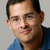
David Schaffer
Professor, University of California, Berkeley -

Lonnie Shea
Steven A. Goldstein Collegiate Professor, University of Michigan -

Brian Aguado
Assistant Professor, University of California, San Diego -
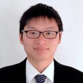
Xiaoping Bao
William K. Luckow Associate Professor, Davidson School of Chemical Engineering, Purdue University -

Shaochen Chen
Professor & Founding Director, University of California, San Diego -

Honggang Cui
Professor, Johns Hopkins University -

Adam Gormley
Associate Professor, Biomedical Engineering, Rutgers University -

Dan Huh
Professor of Bioengineering, University of Pennsylvania -
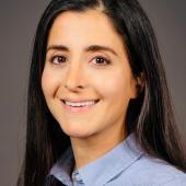
Zeinab Jahed
Assistant Professor of Chemical and Nano Engineering, University of California, San Diego -

Juliane Nguyen
Associate Professor & Vice Chair, University of North Carolina at Chapel Hill -

Sean Palecek
Milton J. and A. Maude Shoemaker Professor of Chemical and Biological Engineering, University of Wisconsin-Madison -
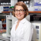
Mana Parast
Professor of Pathology, University of California, San Diego -

Daniel Reker
Assistant Professor, Duke University -

Ankur Singh
Professor, Georgia Institute of Technology -

Yichun Wang
Assistant Professor, University of Notre Dame -
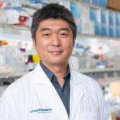
Jun Wu
Associate Professor, University of Texas Southwestern Medical Center -
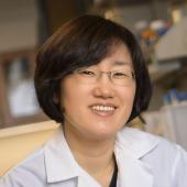
Yoon Yeo
Lillian Barboul Thomas Professor, Purdue University -
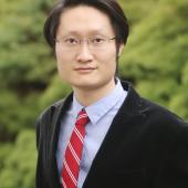
Yu Shrike Zhang
Associate Professor, Harvard Medical School


